A.Y. Chen Illustration & Design
  • Home
  • Portfolio
    • National Science Foundation (NSF)
    • Science magazine art
    • Vector Art
    • Black & White
    • Classic work
  • Animation
  • Blog
  • Contact
  • Children's book art

Shifting into overdrive

6/26/2015

0 Comments

 
Allergic response cascade, antibody binding in allergic reaction, B cells releasing cytokines in allergic reaction, Type I hypersensitivity
© The McGraw-Hill Companies, Inc.
Now comes the action part of the story. Our immune system consists of many different kinds of cells. The important thing to remember is that each cell mainly does two things: signals other cells, and acts upon receiving a signal. The resulting action is quite fast and profound.

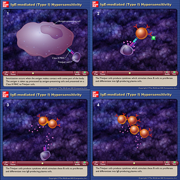
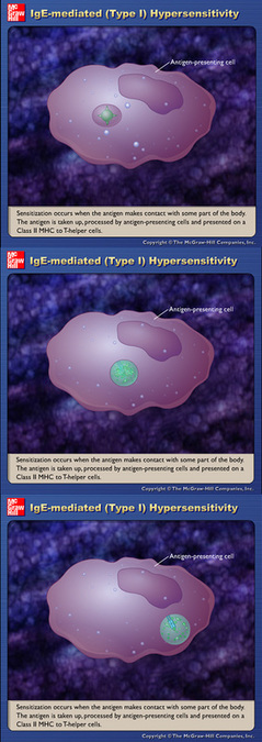
When we last left off in the movie, a presenting cell scavenged our body for things that are not usually present, and grabbed that substance to show a helper T cell a receptor (the signal) on its surface (Lower right corner of frame 1, small plum-colored cell). The helper T cell understands that signal to mean that something bad is in the body, and then communicates with a B cell (frame 2, orange cell), coaxing it to start dividing by releasing cytokines (frames 3 and 4).

There's a lot of terminology and lots of players involved in an allergic reaction. So if you're wondering why (like I was in those very uncomfortable 24 hours following my hives/rash reaction) your medication takes a while to calm down your swelling, itching, and general misery, remember that all these players need to be calmed down as well!
0 Comments

Hey, I know YOU

6/24/2015

0 Comments

 
Continuing on with allergic reactions, this ~2 min. 2D animation from McGraw-Hill Companies, Inc., shows what the static images from the previous post shows:
http://highered.mcgraw-hill.com/olcweb/cgi/pluginpop.cgi…

Let's take each image in turn. The 3 screen captures shown here are taken directly from the animation.

The first step in becoming allergic to a substance is called sensitization. The "antigen" is whatever substance will be that triggers a future histamine reaction (e.g. nuts, cat dander, pollen, bee stings, dust, mold, penicillin).

So, all we're seeing in this sequence is that the irritating substance is being taken up by a specialized cell, whose job it is to translate that irritant into a signal that other cells will understand is NOT GOOD. So in the top image, the green circle with the 4 triangular points within the darker purple bubble-the irritant-is being translated into two objects that look like cattails. The cattails are called receptors, and are what the other cells will recognize as something bad.


Think of it like translating English into another language so that someone will understand what is being said.

Picture
© The McGraw-Hill Companies, Inc.
0 Comments

Haaaaa......CHEW!

6/19/2015

0 Comments

 
© what-when-how.com
What triggers an allergic reaction
Mast cell releasing histamines
© Brycelab.com
We're going to switch gears now and start a series of posts about the basics of an "allergic" reaction. Thanks to the "what-when-how" blog, and the Bryce Lab for these images.

Your body has an amazing line of defense with your immune system, which works with a variety of cells that protect your body from the stuff in the environment that finds its way into you.

But first, it has to determine whether or not the substance is harmful. And for that, it has to be recognized by your body's cellular defense system.

So, the images show the main cell, the mast cell, that is responsible for recognizing the harmful stuff. It then also deploys histamines which bind to specific receptors on other cells. One place H1 receptors are found is on blood vessels. When the histamine binds to that receptor, it makes the vessel more "leaky." This is what promotes fluid leaking into the skin, creating localized swelling and HIVES!
0 Comments

June 17th, 2015

6/17/2015

0 Comments

 
Picture
Although I don't fully understand how the faucium channels are formed to allow the baby to breathe and not choke while drinking, it got me thinking that this ability is a pretty good one, so why do we lose that function when we grow up?
The answer is: when the epiglottis drops down in our throats it allows us to make all kinds of sounds, an important aspect of communication.
So, why DO we choke? Our two pipes for breathing and eating/drinking are both opened to our mouths. The brain involves 4 cranial nerves to automatically start a 3-step process to swallow, one of which is to shut our epiglottis over our airway. But if we are laughing or talking at the same time as food/liquid is in our mouths, we have to breathe. The process of inhaling signals the brain to keep the airway open, and thus food/liquid can accidentally fall into the wrong pipe....thus, we splutter to quickly eject that out of our airway!
0 Comments

It's all about the epiglottis

6/12/2015

0 Comments

 
ERRATA ALERT!
In the previous post, I used a child's anatomy to show how our epiglottis closes over our airway to avoid breathing in what we eat/drink, because there was far less anatomy depicted in this illo than in the adult version. It was easier on the child's head to see the differing positions of the epiglottis, but just to remind you, what you were looking at was not the anatomy of an adult throat.


This leads to an interesting ability that babies possess: have you ever watched a baby drink a bottle for minutes on end, without ever stopping to take in a breath? This is because a baby's throat anatomy is very different from the adult's, in that both the baby's tongue location and the shape of the epiglottis allow for the creation of separate "channels" in order to drink and breathe simultaneously!
For any of you curious about looking into this further, it's called a faucium channel. Anyway, sorry for the potential confusion!

Profile anatomy of an infant's mouth and throat, mouth anatomy of a baby
0 Comments

Talking while eating is not a good idea

6/5/2015

0 Comments

 
Talking about eating and breathing simultaneously (as I often do it) got me to thinking about that amazing piece of anatomy I introduced in the last blog: the epiglottis! This little flap not only works mechanically, but it's also triggered by the brain.

To demonstrate, look at the circled regions in my drawing of a child's head. On the left, the epiglottis is in a position that allows access to both the food/liquid (esophagus) AND the airway (trachea) pipes. On the right, when you swallow, the flap shuts over the opening to the airway, thereby preventing the contents in the mouth from entering the airway!

SO, the question is, can anyone guess how I accidentally on many occasion choke on my food/drink?

Picture
0 Comments


    The purpose of this blog is to explore more effective and exciting ways to communicate science.

    Archives

    June 2017
    May 2017
    April 2017
    March 2017
    February 2017
    January 2017
    December 2016
    November 2016
    October 2016
    September 2016
    August 2016
    July 2016
    June 2016
    May 2016
    April 2016
    March 2016
    February 2016
    January 2016
    December 2015
    November 2015
    October 2015
    September 2015
    August 2015
    July 2015
    June 2015
    May 2015
    April 2015
    March 2015

    RSS Feed

Powered by Create your own unique website with customizable templates.