A.Y. Chen Illustration & Design
  • Home
  • Portfolio
    • National Science Foundation (NSF)
    • Science magazine art
    • Vector Art
    • Black & White
    • Classic work
  • Animation
  • Blog
  • Contact
  • Children's book art

Swelling doesn't make it itch any less

7/31/2015

0 Comments

 
The answer to the last post about the prostaglandin that makes me puff up is: Arachidonic acid! It is found in foods which I love like milk and fish, but might not be the exact reason why my eczema flare-ups occur, nor why say, my mosquito bites tends to be way more dramatic than others.

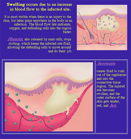
So I'd like to move on to another summer time ailment, at least, for people like me, and that is: mosquito bites! What really interests someone like me who demonstrates a hypersensitivity to the bites, is WHY do the bites swell up so dramatically, and why are they SO itchy that I'd rather scratch myself to the point of bleeding and scarring than leave it alone?

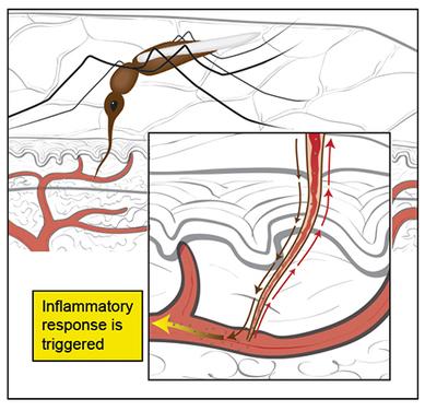
Well, the answer lies in an elegant function of a mosquito's proboscis, its long nose-like instrument that is flexible and thin. It uses this to pierce our skin and then PROBE for a blood vessel, and is so microscopic that we cannot feel it when it goes in. Once it hits the vessel, the object of the game is to suck our blood
up through this proboscis, which is also a straw by the way, that the mosquito needs to feed her eggs. But this instrument can simultaneously eject substances into us that prevent blood from clotting; once blood clots, it can't get sucked up through the proboscis.

A certain protein also found in their saliva, dubbed rAed a 2, is an allergen, or a substance that causes an allergic reaction. We have not isolated, analyzed, nor identified it sufficiently to create a counter punch to it. So for those of us who are sensitive, right now our only hope is to get bitten more and more in order for our bodies to hopefully become de-sensitized to the allergen. Hmmm, I think I'd rather wear DEET!
mosquito bite triggers inflammatory response, mosquito proboscis
0 Comments

Ahhhh, swelling

7/29/2015

0 Comments

 
Inflammation, swelling, increased blood flow, cytokines, prostaglandins
We saw how DNA damaged by UVB radiation is fixed by activating a pathway called NER, which stands for N-ucleotide E-xcision R-epair. This process requires the slowing down of other cellular processes, so can you imagine what would happen when DNA is continually being assaulted by UVB radiation? The continual onslaught of damage causes our DNA to send out an S.O.S.

What are these signals? They are called cytokines, tiny proteins that signal other proteins, or, directly signal a cell to do something helpful. Prostaglandins are bigger molecules that play a major role in the inflammatory response. These signals use our blood stream to travel through to get to the damaged sites. They then squeeze out from the blood vessels into the damaged site, causing that characteristic swelling and pain of sunburn.

Prostaglandins, while extremely important in the inflammatory process, can get a bit overzealous in some folks like me. When they are over-produced they can have negative impacts upon us. Can anyone guess what kinds of negative outcomes can occur?

0 Comments

DNA, heal thyself!

7/24/2015

0 Comments

 
DNA repair mechanism, NER
How does DNA repair itself, that is, swap the C substitutions back out for the correct T pairings?

N.E.R. N-ucleotide E-xcision R-epair.

This is the main pathway, or road map/instructions, that our body uses to repair UVB damage to DNA. This pathway functions to excise, or cut out, the incorrect nucleotides (the C's, or cytosines), and then repair the DNA strand. The steps outlined in the pic have been greatly simplified, in order to just get down the general idea...

DNA damage must first be recognized; this is pretty easy to do as the damaged section of DNA is now a mismatch to its partner strand, because it has the C nucleotides in where there should be T nucleotides. In order to be fixed, the twisted up DNA first has to be unwound in order for the mis-matched pairing section to become accessible. Now, the damaged single-stranded section is cut out. Finally, a new section is made by using the undamaged complement strand as a template by which to make the sequentially-correct complimentary strand.

But what happens if a large number of cells are damaged, as occurs in a severe sunburn? Can all of the cells get repaired? Next post!
0 Comments

It always comes down to DNA

7/22/2015

0 Comments

 
We have to look at our DNA to see what happens to the DNA in our skin cells when exposed to UVB radiation. If you look at the graphic, you'll see that in normal DNA structure, certain compounds, called base pairs, bond together to form the "ladder rungs" of the DNA. A certain purine always pairs with a certain pyrimidine.

When exposed to excessive amounts of UVB radiation, the Thymines get substituted out for Cytosines! And now, there is a defect in the DNA which, if it doesn't get repaired in a timely manner, will lead to the effects we all have experienced as sunburn: burning, red skin, pain, and sometimes, blistering.

To eventually get back to the topic of how disarming this normal (and actually healthy) repair process is linked to a possible treatment for eczema and other skin disorders, we have to skip to another fave topic of mine, the inflammatory response. I'll make the link more explicit in a future post coming soon. But for the next post, I'll continue on with DNA repair!
DNA base pairing, Sunburn damage on DNA level
0 Comments

Summertime fun....???

7/17/2015

0 Comments

 
Sunburn damage on cells, melanocytes, UVA versus UVB
Since we're now into summer, we're switching gears to a new series of posts on some issues we all face in summer time! First up is sunburn, and what is happening on the cellular level.

We all love how the sun feels on our skin, and thus we go out more often when it's warm and sunny. Built within our skin is a protective cell that helps to prevent the "burn," and that is the melanocyte! As you can see from the sketch, this type of cell is found mainly at the bottom of the epidermal layer of our skin; do you see a line of gray, shaded tall skinny cells? That's the bottom of the epidermal layer.

The melanocyte produces the all-important melanin, a substance (pigment) that actually absorbs UV radiation. UV radiation therefore cannot alter/damage other things in its path.
The very interesting part is that when the UV rays actually do their damage to a skin cell, something amazing happens. This in turn has implications for treatment of other skin ailments, and which we'll cover in the next post!
0 Comments

Who's hungry?

7/15/2015

0 Comments

 
Macrophages, foam cells
In our discussions about why mast cells need to be cross-linked by two antibodies per allergen bound, one hypothesis involves the macrophage and IgG density (http://www.sciencedirect.com/science/article/pii/S0165247810001914). A macrophage is the specialized, pacman-like white blood cell. Again, as we've emphasized before, our immune system's cells are pretty specialized, where each responds to different signals that are presented by other cells.

Macrophages are versatile; not only do they help clean up our blood by gobbling up old and dying cells/tissue, but they also have an appetite for things that aren't supposed to BE in our body. Furthermore, they have the ability to take a "piece" of that foreign material, much like an Antigen Presenting Cell (APC), and display it on its surface so that our immune system "marks" it as being bad and knows to react against it in future encounters!

Our pic just shows a collage of macrophages in illustration form and its corresponding "real image" counterpart. Can anyone explain the significance of the macrophage foam cell? The "foam" in the cell is actually lipid, therefore, if a macrophage has been gobbling up lipids, it is an indication of high levels of lipids within the bloodstream. This means that atherosclerosis is likely present. Also, foam cells can indicate certain types of bacterial infections.

Left image: © 2005 Pearson Prentice Hall, Inc.; middle: © 2001 Dennis Kunkel; right: © JAMA.
0 Comments

Mast cell is short for mast cell

7/10/2015

0 Comments

 
mast cells, IgE cross-linking, allergy responders
We've touched upon the antibodies, which are the all-important markers that tell our immune system cells what should be present and what should be destroyed.

Mast cells that have bound to an IgE can now recognize the bad stuff, the allergens. You might have picked up that in a lot of these illos, it's the Ige that's bound to a receptor on the surface of the mast cell that will do the future binding to whatever allergen it's been "sensitized" to. SO, like a lock to a key, that mast cell can now recognize that allergen.

And then that mysterious cross-linking between two IgEs on the mast cell has to occur (see middle image in pic) before the mast cell has the "go-ahead" to release its histamines!

According to the bottom-most diagram, as well as previous posts on this topic, what are two things that histamines will do, AND, how do these actions help the body to rid itself of the allergen?

We'll conclude the topic of allergic reactions with a brief discussion on macrophages.
Top image: © Boston University; middle image: © Elsevier Ltd.; bottom: © 2010, PentaVision, LLC.
0 Comments

Ig is short for Immunoglobulin

7/8/2015

0 Comments

 
Types of antibodies, five types of immunoglobulins, Igs
I posited a question last post asking why two IgE antibody units had to cross-link one molecule of Allergen. No matter how hard I tried, I couldn't find an explanation as to why two IgEs have to be linked when bound to a mast cell's receptors in order for it to be activated. One hypothesis is that it makes the antigen-antibody complex big so it allows phagocytosis, or gobbling up by a large cell called a macrophage, to occur more easily.

To be more comprehensive, I'm going to expand upon immunoglobulins and mast cells.

An immunoglobulin (Ig), also called an antibody, is a Y-shaped molecule that binds to anything foreign in our bodies. In this way, the Ig marks what it binds to for other cells to look at and destroy. IgE is the least common of the 5 different kinds of immunoglobulins, unless you're allergy-prone, like me!

In this pic, you'll see how each type of Ig is structured differently, but made from an identically-shaped repeating structure. Keep in mind that a molecule's shape is extremely important in chemistry, and that the principle that each object binds to another object in a very specific way, holds true. So, if these Igs have different shapes, you can assume that they will bind to different structures in very specific ways. Likewise, if their shape is similar (as with the three in the top row), they may be able to bind to similar or the same type structure on another cell.
For an additional description of each type of Ig, where they are found, and what they do, see: http://www.webmd.com/a-to-z-guides/immunoglobulins
© Nature Reviews/Molecular Cell Biology
0 Comments

The finale: Sneezing, itching, watery eyes and general misery

7/3/2015

0 Comments

 
From the previous post, I asked two questions. And so, in answer to those questions, the 5th cell is called a mast cell (frame 1) and the 4th signal is an antibody.

The cascading pattern of one cell triggering an action in another cell continues with this mast cell, but with one important difference: for an action to be triggered, two of the red Y-shaped bound antibodies, called IgEs (Ig short for immunoglobulin), have to bind to one molecule of the bad stuff (green spiked spheres in frame 2). Can ANYONE explain why this is the case?

Once this occurs, the mast cell releases histamine (cyan blue particles, frame 3), which triggers your nose to run, the swelling, the itching, and the sneezing. All the hallmarks of allergies.

Another really important note is that these allergic responses occur the SECOND time you are exposed to the bad stuff. The FIRST time is like the practice run, where the cells learn that the bad stuff IS bad. Our immune system is so smart that the red, Y-shaped IgEs that do the "recognizing" stay in your body for weeks, as if they are anticipating that if the bad stuff could get in once, it'll get in again, very soon after.

It may seem crazy that we are made to be so miserable with our allergic responses, but without the immune system, we would be in far worse shape!

Allergic response cascade, antibody binding in allergic reaction, B cells releasing cytokines in allergic reaction, Type I hypersensitivity
© The McGraw-Hill Companies, Inc.
0 Comments

July 03rd, 2015

7/3/2015

0 Comments

 
0 Comments
<<Previous


    The purpose of this blog is to explore more effective and exciting ways to communicate science.

    Archives

    June 2017
    May 2017
    April 2017
    March 2017
    February 2017
    January 2017
    December 2016
    November 2016
    October 2016
    September 2016
    August 2016
    July 2016
    June 2016
    May 2016
    April 2016
    March 2016
    February 2016
    January 2016
    December 2015
    November 2015
    October 2015
    September 2015
    August 2015
    July 2015
    June 2015
    May 2015
    April 2015
    March 2015

    RSS Feed

Powered by Create your own unique website with customizable templates.