 | 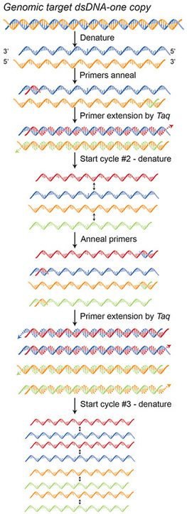 |
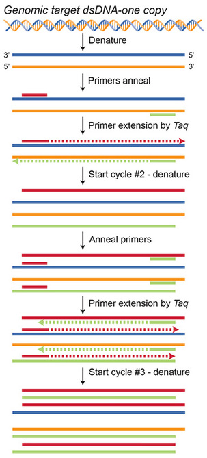
The left image is the same one shown in the last post, only now with labels. In the right image I use the actual structure of DNA, rather than the simple colored lines, to represent the two strands that make up one whole strand of DNA. Here you can use the color coding to understand how these two images correlate.
The very simplified explanation of how DNA copies itself is that one whole DNA strand, made of one blue and one orange strand, is first unwound and then separated. Special pieces of DNA called primers–the short red and green lines–stick (anneal) to each unwound DNA strand, because each is designed to be the compliment base pair sequence as that stretch of DNA. Taq, the enzyme, can now stick to the primer and read the DNA sequence in the direction of the arrow, adding each compliment base pair to the end of the primer in a chain-like manner. Therefore, the beginning of the new strand starts with and incorporates the primer.
Now, can anyone tell me what's happening in cycle #2?
 RSS Feed
